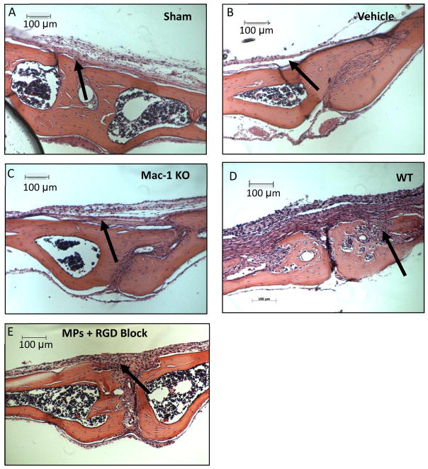
Figure 8.
Decalcified calvaria were sectioned and stained with hematoxylin and eosin for osteolysis measurement. Representative images of calvaria from A) Sham and B) Vehicle C) Mac-1 KO D) WT and E) WT + RGD loaded EVA disc are shown to depict the difference in area of osteolysis, with regions of osteolysis indicated (arrows).

