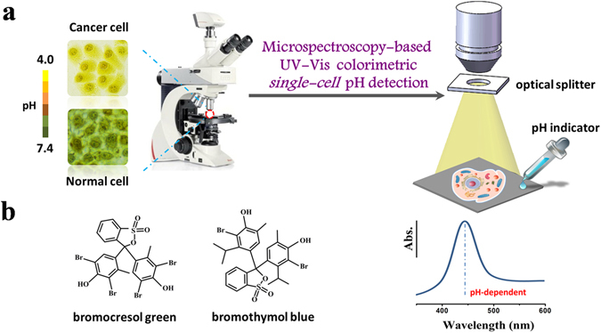
Figure 1.

Schematic of colorimetric single-cell pH imaging and detection with two pH indicators. (a) Schematic of single-cell pH imaging and detection by combined use of bright-field microscope-based UV-Vis microspectroscopy and common pH indicators. A selected area single-cell absorption spectrum is collected by an optical microscope equipped with a portable spectrometer and an optical splitter with a small collecting area. (b) The structures of two pH indicators used in this study.
