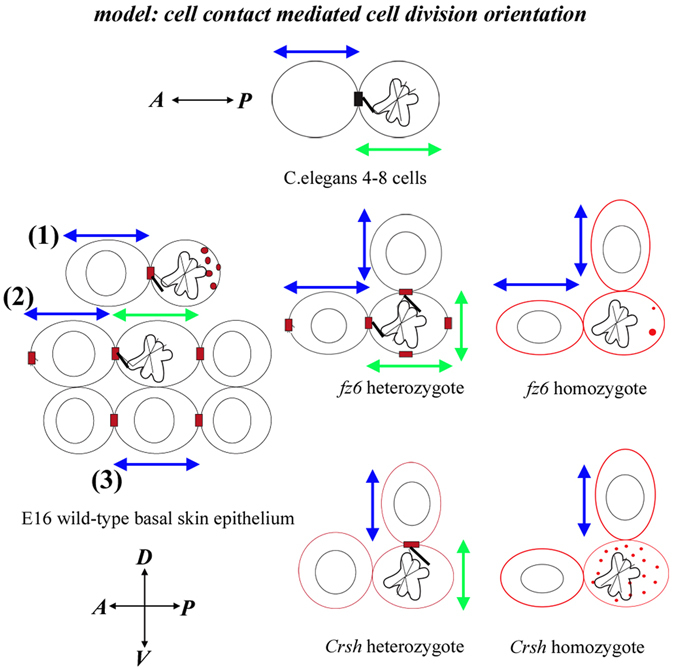
Figure 9.

Model of core protein dependent cell-contact mediated orientation of cell division in the mammalian skin. Taken together our data suggest that in wild-type the asymmetric cell surface cue which aligns cell division plane with neighbouring interphase long axis extent can be uni-polar (enriched at the interface between mitotic cell and the interphase neighbour) as shown in division 1 or bi-polar as shown in division 2,3. The asymmetric cue can be located ‘head-on’ with the mitotic cell as shown in division 1 and 2 or alongside the mitotic cell as shown in division 3. A: anterior, P: posterior, D; dorsal V; ventral. Blue arrows denote interphase long axis orientation, green arrows denote PCD orientation. In C. elegans cartoon, black box labels Wnt/Frizzled cell surface cue. Red boxes label Celsr1/Fz6 cell surface asymmetry. Black lines denote putative link between the cell contact cue and the mitotic spindle. Red dots denote Celsr1/Fz6-expressing intracellular vesicles. In core protein mutants Celsr1 planar polarised asymmetry during interphase diminishes as marked by red cell outlines.
