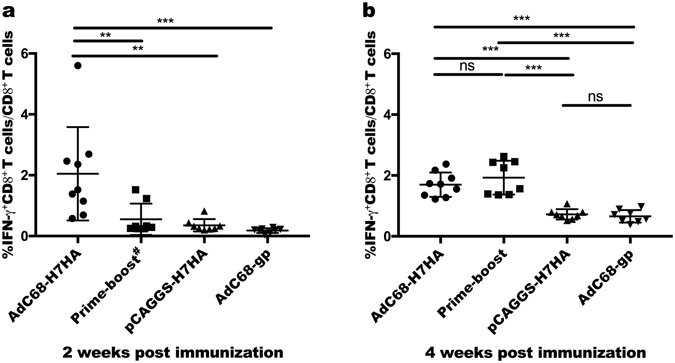
Figure 2.

H7N9 virus-specific T cells responses. Two and 4 weeks after immunization, mouse PBMCs (peripheral blood mononuclear cells) were separated and collected. PBMCs were stimulated with H7HA peptide pools (10 μg/mL) 2 hours before adding GolgiPlugTM for another 4 hours of incubation. The percentage of CD8+ T cells secreting IFN-γ was then measured by intracellular cytokine staining. (a) H7N9 virus-specific T cell responses at 2 weeks after prime immunization. (b) H7N9 virus-specific T cell responses at 4 weeks after prime immunization. The error bars represent the SD. ***p < 0.001; **p < 0.01; *p < 0.05. #At 2 weeks post immunization, the prime-boost group received DNA priming, as in the DNA-only group.
