
Figure 4.
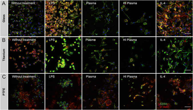
Immune-histological staining of intercellular adhesion molecule CD54 (green) and β-Actin (red) illustrated morphological changes in dependency of test conditions and material on (A) glass, (B) titanium and (C) PTFE. An uneven topography and a long working distance prohibited to capture sharp images on silicone and PE surfaces. The scale bar depicts 50 µm and is valid for all images. The following abbreviations are used: HI for heat-inactivated.