
Figure 4.
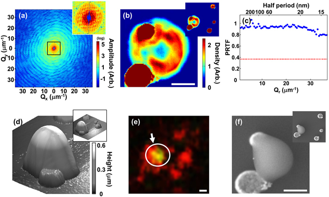
(a) Diffraction amplitude from a specimen with a mitochondrion together with Au reference objects. The central part is enlarged in an inset to display details of the speckle pattern. (b) CXDI projection image of the mitochondrion reconstructed from (a). The red circular objects are the Au reference objects. An image with an enlarged field of view is included in the upper right corner. (c) PRTF evaluated from the iterative reconstruction which indicates that the image resolution is around 14 nm. Red dotted line indicates 1/e, the resolution criterion. (d–f) AFM (d), fluorescence (e), and SEM (f) image of the same specimen. The green fluorescence emission indicated in (e) shows that the measured object is indeed a mitochondrion. The scale bars are 400 nm.