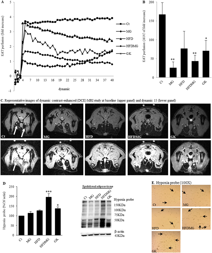
Figure 2.

Glycation impairs pEAT blood flow and causes hypoxia in adipose tissue. Blood flow was evaluated through the area under the curve (B) of a dynamic contrast-enhanced (DCE) MRI study (A). Representative images at baseline and dynamic 15 (C). Hypoxia was assessed using an antibody against pimonidazole adducts by WB quantification (D) and IHC (E). Ct - Wistar 12 m; MG - Wistar + MG supplementation; HFD - HF diet-fed Wistar; HFDMG - HF diet-fed Wistar + MG supplementation; GK - Goto-Kakizaki 12 m. Bars represent means ± SEM, n = 6–8. * vs Ct. 1 symbol p < 0.05; 2 symbols p < 0.01; 3 symbols p < 0.001.
