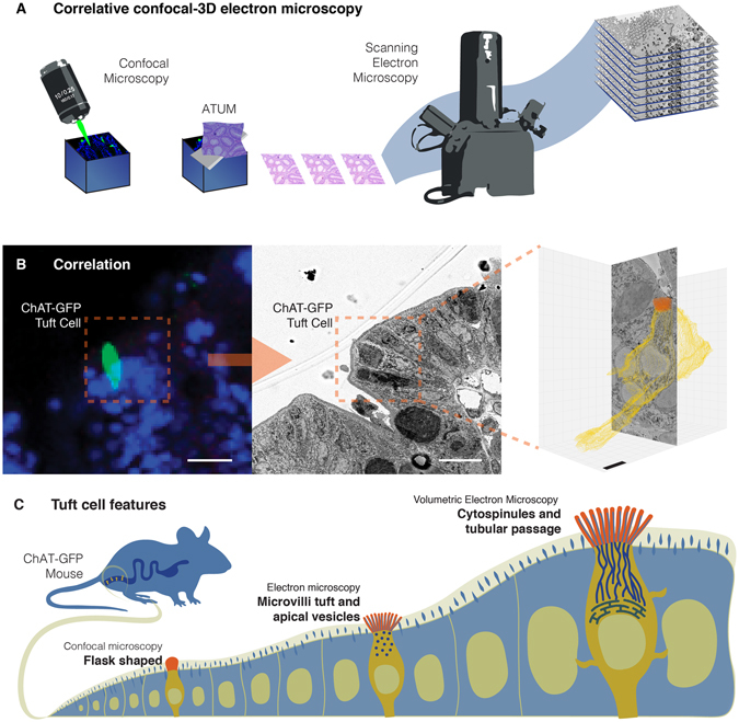
Figure 1.

Identifying tuft cells for volumetric electron microscopy. (A) Overview of correlative method to identify a specific cell by fluorescence, then performing targeted scanning electron microscopy to uncover the ultrastructure of the desired cell in the third dimension. (B) A tuft cell in the colonic epithelium of ChAT-GFP transgenic mice is identified by fluorescence (B-left) and ATUM SEM (B-center), then volume rendered from serial images using data visualization software (B-right). (C) Volumetric EM analysis of tuft cells revealed cytospinules and a gut-to-endoplasmic reticulum passage. Bars in B-left and middle = 10 µm, bar in B-right = 1 µm.
