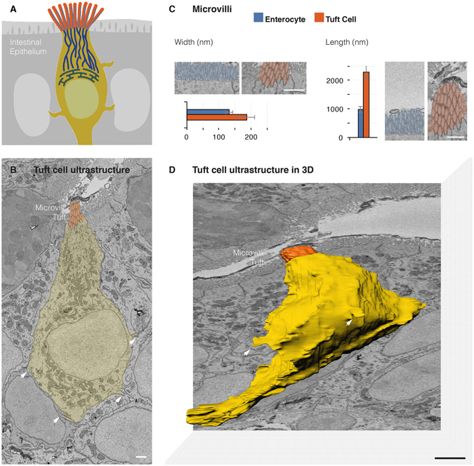
Figure 2.

The ultrastructure of the tuft cell in the third dimension. (A) Schematic overview of the intestinal tuft cell. All data in panels are oriented from top-lumen to bottom-basal lamina. (B) An SBEM micrograph revealing a tuft cell in the mouse distal small intestine (yellow). Cytoplasmic spinules (arrows) projecting into adjacent cells and vesicles beneath the microvilli are evident at this resolution (5 nm/pixel). (C) Morphometrical assessment of tuft cell microvilli. Measurements are an average of 30 individual microvilli from three different cells in each group. (D) Volume rendering of 500 consecutive SBEM micrographs shows the complete 3D ultrastructure of tuft cell in the third dimension, including microvilli (orange), basal process, and cytospinules (arrows). See also Video 1. Bars = 1 µm.
