
Figure 4.
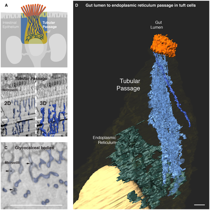
A molecular passage to the endoplasmic reticulum. (A) An overview of a passage in the tuft cell connecting its endoplasmic reticulum to the gut lumen. (B) A continuous tubular passage starting at the base of the tuft cell microvilli (left) becomes evident after volume rendering of ATUM images (right). (B) Glycocalceal bodies (arrows) were observed within microvilli as well as within the tubular passage. (D) Volume rendering of ATUM images reveals a passage in tuft cells. It is formed by tubules that start at the base of the microvilli and extend to the endoplasmic reticulum. The tubules appear to merge with the endoplasmic reticulum but this cannot be discerned at the current resolution (4 nm/pixel). See also Video 2. Bars = 1 µm.