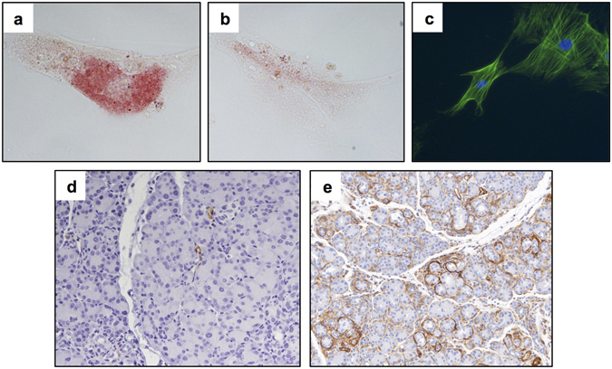
Figure 1.

PSC display an activated phenotype in culture and in a murine model of CP. PaSC were treated with (a) 10 μM all-trans retinoic acid (ATRA) or (b) vehicle control for 48 hours and stained for Oil-Red O. Cells were analyzed by light microscopy at 40X magnification. (c) Untreated PaSC were stained for α-SMA (green) by fluorescent microscopy following 48 hours of incubation (DAPI counterstain, 40X magnification). (d) Formalin fixed paraffin embedded (FFPE) pancreatic tissue from mice with caerulein-induced pancreatitis after (d) 1 week and (e) 5 weeks of treatment were stained for α-SMA (20X magnification). Representative images from n = 5 mice per group.
