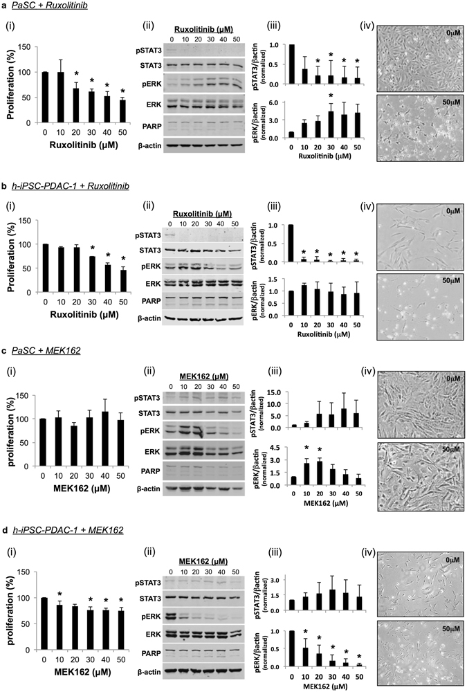
Figure 3.

Effect of Jak/STAT and MEK inhibition on PSC proliferation in vitro. (a,c) PaSC or (b,d) h-iPSC-PDAC-1 were treated with ruxolitinib (a,b) or MEK162 (c,d). (i) After 24 hours of incubation, cell proliferation was analyzed by MTT assay. (ii) Lysates were also taken at 24 hours and analyzed by western blot. β-actin served as a loading control. (iii) Results were quantified via densitometry and normalized to β-actin. Graphs display mean ± STD from 3 biological replicates (* indicates p < 0.05). (iv) Light microscopy images were taken of treated cells following 72 hours of incubation (40X magnification).
