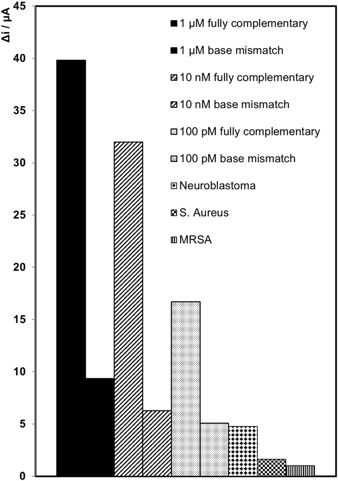
Figure 5.

Comparison of the change in current for the fully complementary target strand (miR-134), the one base mismatch (miR-758) target strand and fully non-complimentary neuroblastoma (black diamonds), S. aureus (black squares), and MRSA (black vertical lines) nucleic acid strands. Three different target concentrations were used for miR-758; 1 µM (solid black), 10 nM (black lines) and 100 pM (black dots). Concentration of the miR-132-3p, SA, and MRSA nucleic acid target strands are 1 µM. Concentration of capture and probe miRNA strands are 1 µM, where the probe miRNA strand is labelled with electrocatalytic PtNPs. Concentration of H2O2 added is 20 µM. Potential applied is −0.25 V in 1 mM DPBS.
