
Figure 4.
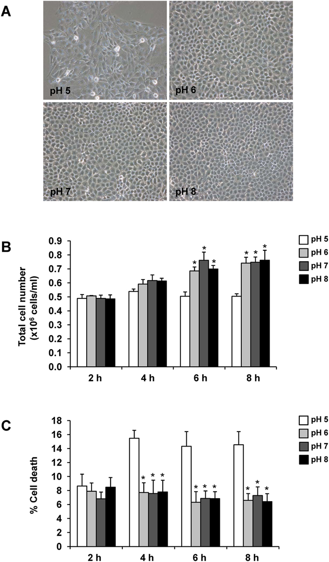
Effect of pH on renal tubular cell proliferation and death. MDCK cells were cultivated and maintained in medium with different pH (from 5.0 to 8.0) for up to 8 h. (A) Cell morphology observed under a phase contrast microscope at 8 h with original magnification of 200×. (B) Total cell number representing cell proliferation. (C) Cell death assay using Trypan blue staining. The data are reported as mean ± SEM of the data obtained from 3 independent experiments. *p < 0.05 vs. pH 5.