
Figure 1.
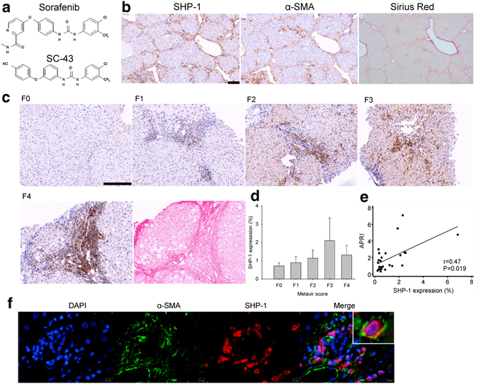
SHP-1 phosphatase is associated with liver fibrosis. (a) The structure of sorafenib and its derivative SC-43. (b) SHP-1 located primarily in areas with α-SMA-expressed activated HSCs and significant fibrosis by sirius red staining of the CCl4-induced fibrosis mouse liver. (c) SHP-1 overexpression in fibrotic areas of patients with CHB with advanced fibrosis, graded by the Metavir scores (F0–F4). The SHP-1 located in sirius red-positive fibrotic areas. (d) SHP-1 expression according to Metavir score. (e) SHP-1 expression positively correlated with APRI. (f) SHP-1 colocalized with α-SMA-expressed activated HSCs in human liver. Scale bar: 200 μm.