
Figure 2.
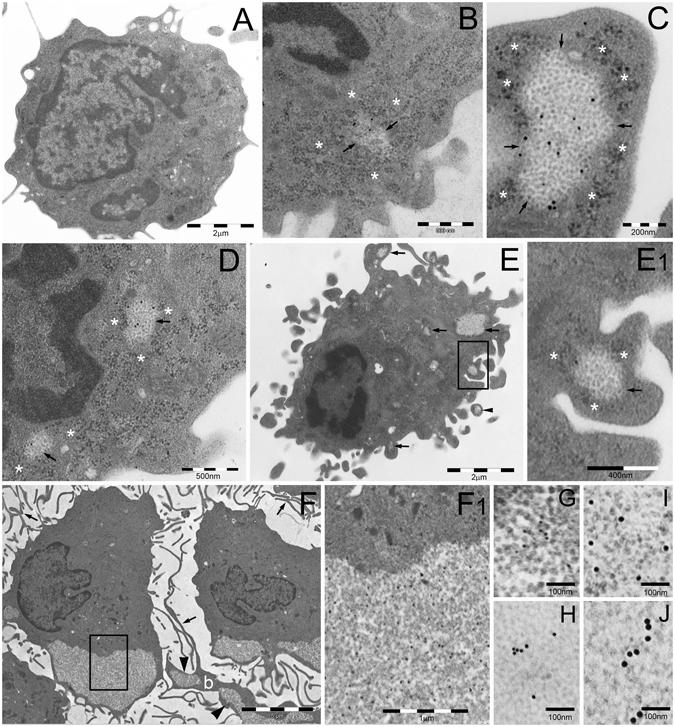
TEM analysis of PaCS development in IL4-DC. (A) Untreated human blood monocyte showing moderately developed cytoplasm with only a few organelles and cell protrusions. Cells treated with GM-CSF plus IL-4 for 24 h (B,C) or 48 h (D,E) showing early PaCS (arrowed borders) development inside ribosome-rich cytoplasmic areas in the absence of ER cisternae. Note FK1-reactive polyubiquitinated proteins (B,E) and 20 S (C,D) immunogold deposits in PaCS formed by moderately electron dense, barrel-like particles, to be compared with more dense and irregular, PaCS-sorrounding, ribosome particles (asterisks). The cell in (E) shows abundant cytoplasm with short hobnail-like protrusions and scattered PaCS (arrows); one of which (boxed) inside a protrusion is enlarged in (E1). Also note in (E) a detached PaCS-storing vesicle (arrowhead). Large PaCS are seen in DC after 5 days treatment with GM-CSF plus IL-4 (F boxed area enlarged in F1 to show PaCS barrel-like particles with selective 20S immunogold reactivity). Note in (F) long and thin cell protrusions (arrows) typical of well-differentiated DC and a long bleb (b) filled with PaCS (arrowheads). 19S proteasome (G 10 nm gold particles), HSP40 (H, 15 nm), HSP70 (I, 20 nm) and HSP90 (J, 20 nm) immunoreactivity was found in enlarged portions of PaCS taken from sections adjacent to (F).