Figure 3.

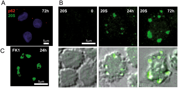
Confocal microscopy of PaCS in IL4-DC. (A) IL4-DC incubated for 72 h with GM-CSF plus IL4, processed according to the common sample preparation for confocal microscopy procedure and immunostained with proteasome 20S (green) and p62 (red) antibodies. No positive 20S immunofluorescent structures were visible, while a few small p62-storing fluorescent bodies (red), likely corresponding to autophagosomes, appeared in some cells. (B) IL4-DC incubated for 0, 24 or 72 h, fixed, and processed as for TEM and immunostained for 20S proteasome (green). Proteasome-reactive PaCS were visible after 24 and 72 h incubation, but not in control, untreated monocytes. Corresponding phase-contrast images are shown below to help identify the cells. (C) Immunofluorescent PaCS were also seen in IL4-DC after 72 h incubation, after immunostaining for polyubiquitinated proteins with FK1 antibody.
