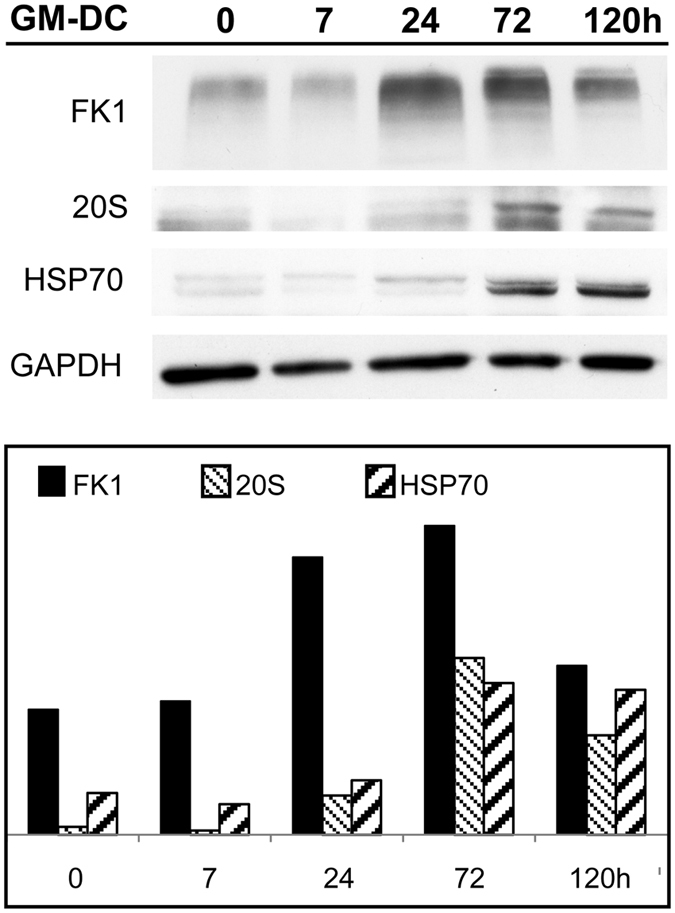
Figure 4.

Biochemical evidence of PaCS development. Immunoblotting of DC lysate at various stages of differentiation from their peripheral blood monocyte precursors treated with GM-CSF plus IL-4 for 0–5 days. The histogram shows FK1, 20S and Hsp70 quantitation normalized for protein loading (GAPDH). Note the increment in the levels of FK1-reactive polyubiquitinated proteins and 20S proteasomes starting from 24 h and that of Hsp70 starting from day 3.
