
Figure 5.
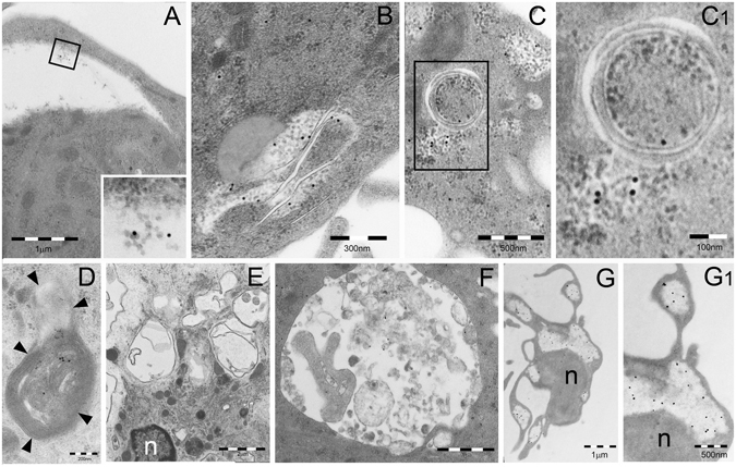
TEM analysis of PaCS autophagy and bleb formation in IL4-DC after IL-4 withdrawal. TEM of cells incubated for 5 days with GM-CSF plus IL-4 and then for 3 (A–D) or 5 (E–G) days in RPMI–FBS without cytokines. (A) A clear area, not delimited by membrane, was seen in the cytoplasm of a DC. This was identified as an empty PaCS, due to its residual barrel-like particles and 20S immunogold reactivity, visible at higher magnification in the inset. (B–D) Autophagic double or multilayered membranes penetrate and wrap/envelope cytoplasmic areas containing FK1-positive PaCS. See structural details in the enlargement (C1) and p62 immunoreactivity of an autophagosomal vesicle with fused multilayered membranes in (D). (E–G) A collection of empty autophagic vesicles and no PaCS are seen in (E), while in (F) a single-membrane vesicle shows heterogeneous, lysosome-type dense bodies and sparse, residual immunogold reactivity for the autophagic marker LC3. (G) FK1-reactive PaCS were still recognized in the cytoplasm as well as in the cell blebs, although most barrel-like particles were dissolved. Nuclear and cytoplasmic densification, with loss of details and organelles, were indicative of ongoing apoptosis. See enlargement in (G1). n, nucleus.