
Figure 6.
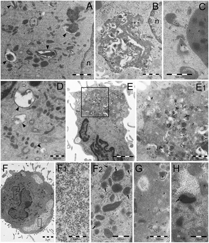
TEM analysis of DALIS development in GM/LPS-DC (A–C) or IFNα-DC (D–E). (A) Numerous endosomes of various shape and density and autophagic vesicles (arrowheads) are scattered in the cytoplasm of a DC incubated with GM-CSF for 5 days. (B) In cells first incubated with GM-CSF as in (A) and then treated with LPS, DALIS appear formed by a collection of vesicles with interposition of amorphous material and dense bodies. DALIS likely result from aggregation of autophagic vesicles and endosomes, as suggested in (C) by the LC3-reactive endosomes contacting a DALIS (on the right). (D) Abundant endosomal structures and few autophagic vesicles (arrowheads) are also found in the cytoplasm of a DC cell treated for 3 days with GM-CSF plus IFNα. (E) A large DALIS in the cytoplasm of another cell, treated as in (D), which is enlarged in (E1) to show vesicles and dense bodies embedded in an amorphous component. (F) An LPS treated IL4-DC showing PaCS (enlarged in F1) and aggregated endosomes (enlarged in F2). PaCS are negative for K63-linked polyubiquitin chains antibody (F1) while some endosomes (arrowheads) (F2) as well as a DALIS (G, from an adjacent cell) show a clear reactivity. (H) The enlarged cytoplasm of another cell treated as in (F) and (G) shows p62 reactivity of an endosome (arrow) and lack of reactivity of an adjacent PaCs. m, mitochondria.