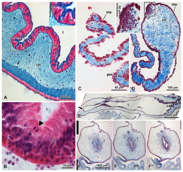
Figure 7.4.
Histological organization of the digestive tube of the sea cucumber H. glaberrima in noneviscerated animals (A) and (B) and at different time points of regeneration (C–F″). (A) Cross-section of the intestine in a noneviscerated animal stained with alcian blue (pH 2.5) and nuclear fast red. The inset shows a higher magnification view of the luminal epithelium. (B) Dividing cell (anaphase, arrowhead) in the luminal epithelium of the normal intestine stained with hematoxylin and eosin. (C–F″) Regenerating digestive tube stained with Heindenhein’s azan. (C and D) Cross-sections through the early gut rudiment on days 3 and 7 postevisceration, respectively. The inset in D shows a detailed view of the mesothelial cells ingressing into the underlying connective tissue. (E) Longitudinal section through the growing gut rudiment on day 14 postevisceration. The growing tip (arrow) is on the left, whereas the cloacal stump is on the right. (F–F″) Day 14 postevisceration; successive cross-sections of a posterior gut rudiment taken through the growing tip (F) and at distances of 40 μm (F′) and 110 μm (F″). cs, cloacal stump; ctl, connective tissue layer of the gut wall; l, gut lumen; le, luminal (mucosal) epithelium; me, mesothelium; pm, proximal mesentery.

