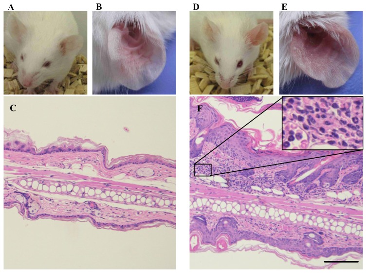
Figure 2.
Typical phenotypic images, close-ups, and histological images of left ear on Day 5. (A, B, C) Normal control animals received neither IMQ nor vehicle cream. (D, E, F) IMQ application on left ear skin. F, inset, a close-up image of inflammatory cells in dermis. Ear thickening, erythema, and scaling are observed on gross appearance. The histological image shows parakeratosis, hyperkeratosis, acanthosis, infiltration of lymphocytes and neutrophils in thickened dermis. Scale bar = 100 μm

