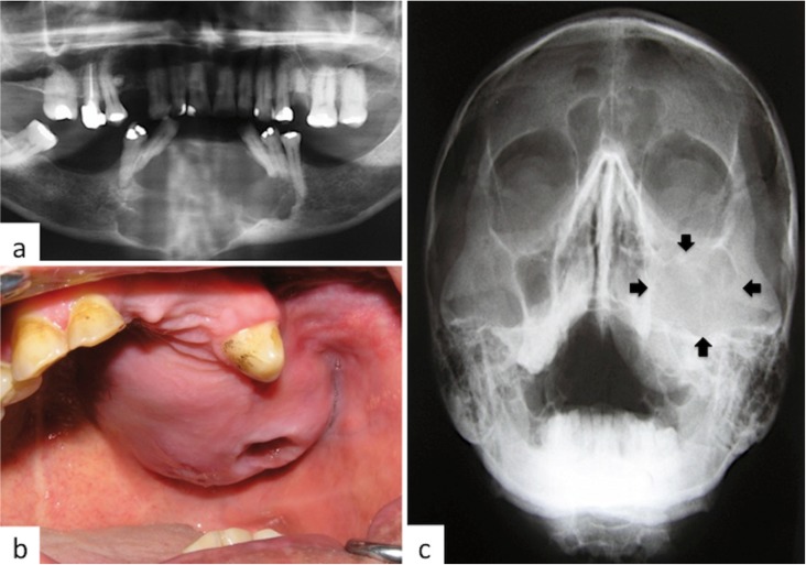
Figure 2.
Most cases of ameloblastic carcinoma involved the posterior mandible. This figure illustrates two unusual cases, one involving the anterior mandible and the other the left maxilla, a. Panoramic radiography showing irregular radiolucent lesion involving the anterior mandible causing tooth displacement, mobility and loss, b. AC of left maxilla affecting the alveolar ridge and extending into the palate. The perforation in the mucosa corresponds to a recent tooth extraction, c. Radiograph of the same case showing radiolucent image involving the left maxillary sinus and adjacent areas, including floor of the orbit.

