Figure 3.

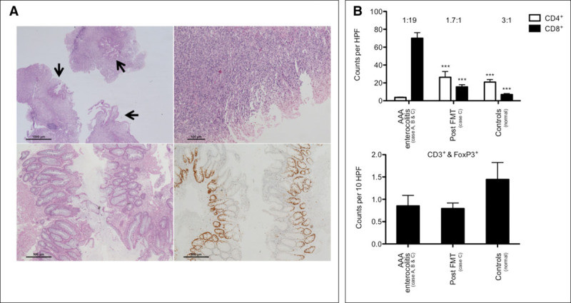
Histopathology and mucosal immunophenotype pre- and post fecal microbiota transplantation (FMT). (A) Colon histopathology 12 d ahead (top left) and at the day of FMT (top right). Only focal residual epithelium is present (arrows). Seven days after FMT, the epithelial lining is reestablished (bottom left). Ki-67 immunohistochemistry identifies proliferating crypt epithelia (bottom right). (B) Significantly increased CD8+ T cells and significantly reduced CD4+ T cells during acute disease in the colon, this immunophenotype is reversible after FMT (top); CD4-to-CD8 ratio is given above bars (one-way analysis of variance [ANOVA], Bonferroni-corrected, *** p < 0.0001). CD3+FoxP3+ double-positive Tregs are not significantly altered (bottom; one-way ANOVA, p = 0.2801). AAA = antibiotic-associated apoptotic, HPF = high power fields.
