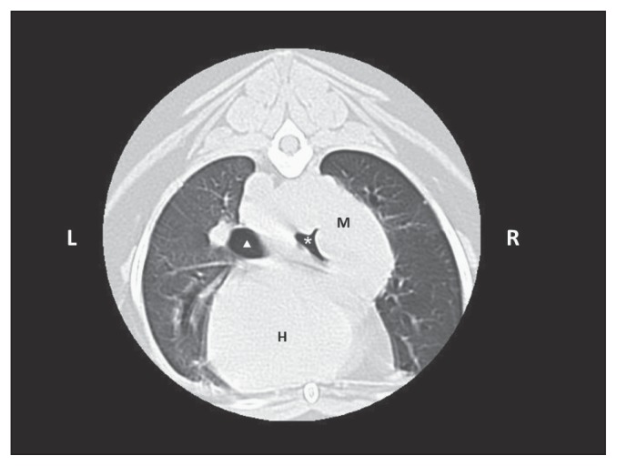
Figure 4.
Computed tomographic image of the dog of Case 2 at the level of the 6th rib, caudally to bronchi bifurcation. Note the mass (M) in the caudal right lobe, dorsal to the heart (H), associated with a severe compression of the main right bronchus (white asterisk) and a mild compression of the left main bronchus (white triangle).

