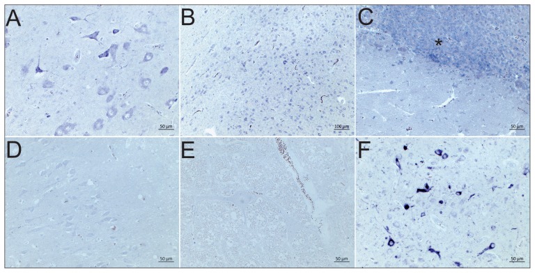
Figure 1.
Detection of BoAstV CH13/NeuroS1 in brain sections by RNA in-situ hybridization. A — Case 23, midbrain and B — case 31, cerebral cortex with strong dark purple positive labeling of neurons; C — Case 37, cerebellum; the granular layer shows a strong neuronal positive RNA labelling in almost all granular cells (asterisk); D — BoAstV Ch13/NeuroS1 negative case 8, brainstem; E — BoAstV CH13/NeuroS1 negative control; F — BoAstV CH13/NeuroS1 positive control.

