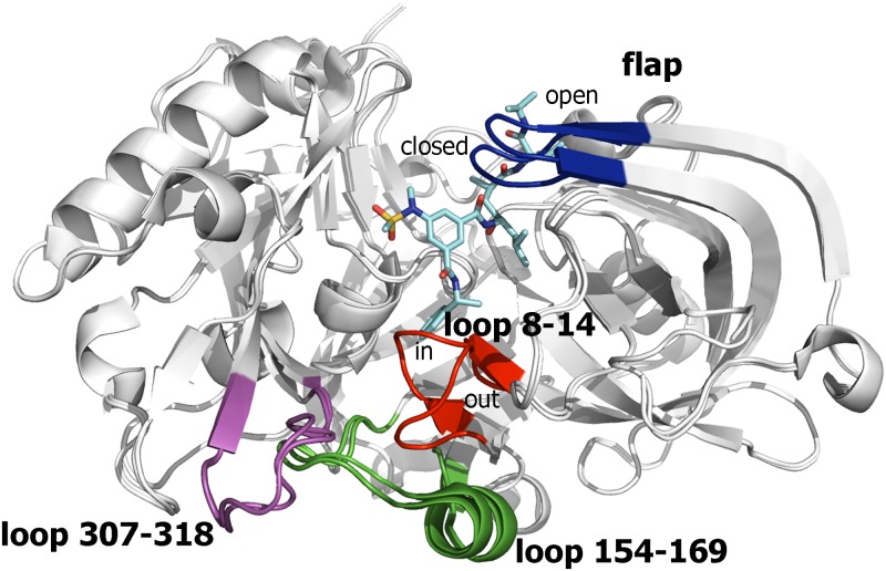
Fig 3. Representation of BACE-1 as found in the structures 1SGZ and 2P4J.
The backbone of the superposed proteins is shown as white cartoon, and the ligand located along the catalytic cleft in 2P4J is shown as cyan-colored sticks. The flap region (blue) shows the open and closed conformations typically found in apo and substrate-bound states, respectively. The other flexible region is located in a region distal from the catalytic site and is shaped by residues 8–14 (red), 154–169 (green), and 307–318 (magenta). The two major conformations found for the loop formed by residues 8–14 is shown. Loops 154–169 and 307–318 are generally disordered in other PDB structures.

