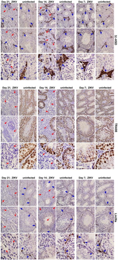
Extended Data Figure 2. Temporal loss of cellularity in the testis after ZIKV infection.

Seven week-old WT C57BL/6 mice were treated with 0.5 mg of anti-Ifnar1 at day -1 prior to subcutaneous inoculation of mouse-adapted ZIKV Dakar. Immunohistochemical analysis was performed on testis tissues collected from uninfected (top panels) or ZIKV-infected animals (days 7, 14 or 21 after infection; bottom panels) at 20× (left two images) and 40× (right image) magnification. Staining was performed with antibodies against 3β-HSD (Leydig cells, left panels), TRA98 (germ cells, middle panels), and Lin28a (type A undifferentiated and type B spermatogonia, right panels). Blue arrows indicate staining of Leydig cells (left panels), germ cells (middle panels), and spermatogonial stem cells (right panels). Red arrows indicate areas of virus-induced damage and loss of tissue integrity and specific cellularity. Scale bars = 200, 200, and 50 μm for the grouping of the three sets of images.
