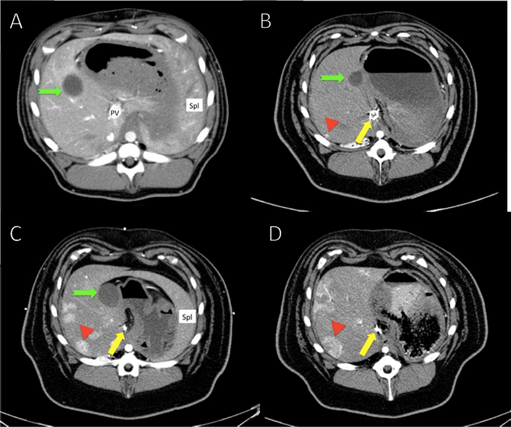
Fig. 1. Computed tomography studies of hepatocellular carcinoma (HCC) tumors in a representative diethylnitrosamine (DEN)-treated Yucatan miniature pig.
Following embolization and treatment with DEN, the pigs developed focal hypervascular lesions in multiple areas throughout the liver as shown by contrast enhancement (red arrowhead). In keeping with the progression of neoplastic lesions, these lesions culminated in the appearance of nodules, which were histologically shown to be HCC.(A) Baseline scan. During all phases of the initial scan, liver parenchyma enhanced evenly. The portal vein was readily identified on the first scan before embolization but was not well visualized on subsequent studies, particularly in the embolized region. (B) Scan at 16 months shows slight medial shift of the gallbladder owing to the shrinkage of the embolized left side of the liver (green notched arrow). The coils in the portal vein (yellow arrow) and the tumor (red arrowhead) are indicated. (C) Scan at 22 months. Note growth in both size and degree of enhancement of the index lesion and the background of additional similar lesions progressing over time until (D) final scan at 28 months just before the pig was euthanized.

