
Figure 2. IBC 3D tumor spheroid high content assay.
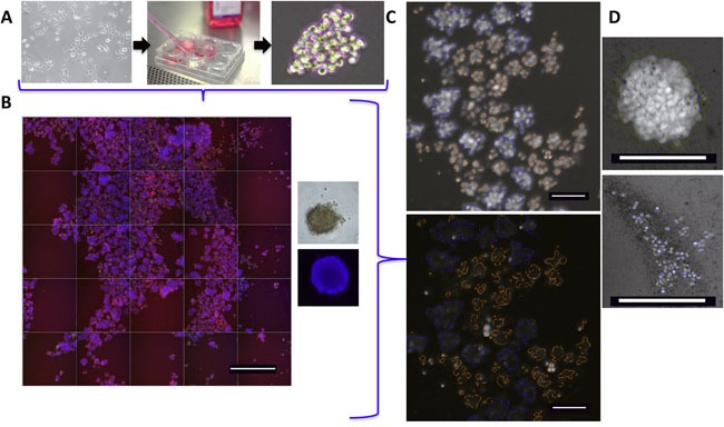
A. Schema of IBC tumor spheroid culture in ultra-low attachment plates and analysis after addition of specific dyes. B. Representative montage of 25 neighboring fields from a single well (10x – scale bar = 800 microns) of spheroids stained with Hoechst 33342, YOYO-1 and MitoTracker Red FM dyes. Right - Enlarged image of a single SUM149 tumor cell cluster shown (phase contrast and Hoechst stained). C. Representative image analysis (10x – scale bar = 100 microns) of SUM149 spheroids at day 6, with masks overlaid over the spheroids (top) or masks alone (bottom) established by Hoechst signal and a >50 μm area cutoff. These regions of interest are then used for analysis of spheroid YOYO-1 and MitoTracker staining. D. Image of a single spheroid (top - untreated and bottom - treated with a cell death inducing agent) to highlight the spheroid mask (blue outline) and morphology.