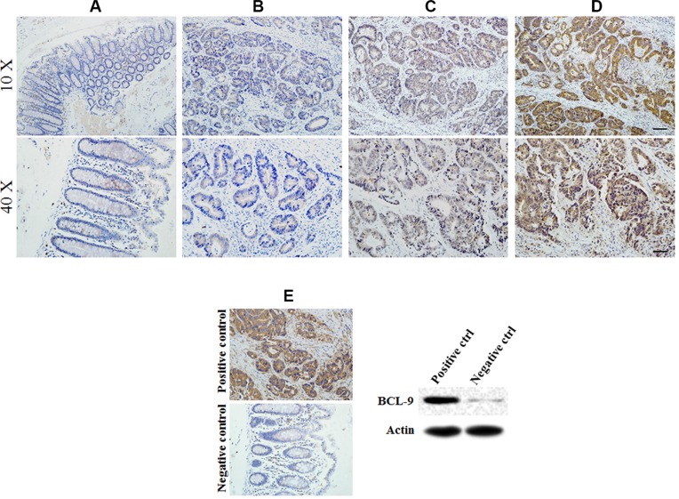
Figure 1. BCL-9 expression in different developing stages of colorectal cancer tissue.
Representative immunohistochemical stained imaging of BCL-9 expression in different developing stages of colorectal cancer tissue containing 284 cases were shown. Positive cells were stained brown. (A) BCL-9 expression in normal mucosa. (B) low intensity expression in colorectal cancer cells scored as “−”. (C) moderate intensity expression in colorectal cancer cells scored as “+”. (D). high intensity expression in colorectal cancer cells scored as “++”. (E) Positive and negative control of BCL-9 expression showed the specificity of staining and were validated by Western-blot assays. Scale bar: 40 μm for low magnitude images (10×); 10 μm for high magnitude images (40×).

