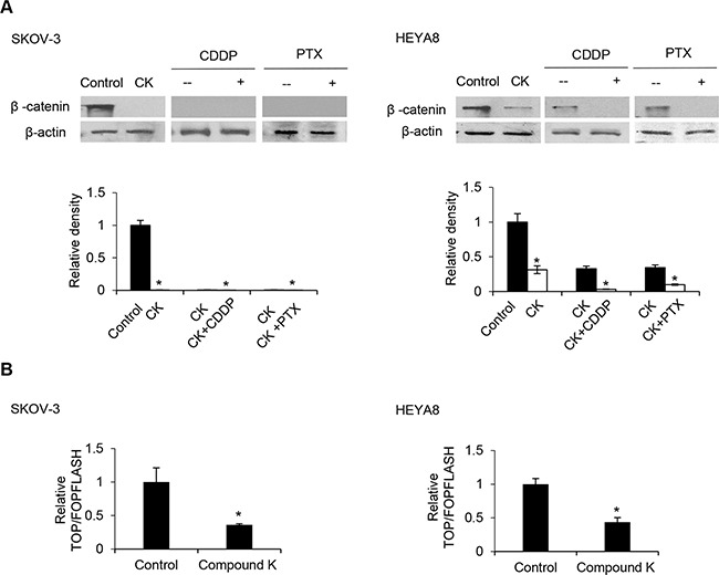
Figure 7. Compound K enhances the chemosensitizing effect through the β-catenin/TCF signaling pathway.

A. Protein levels of β-catenin were analyzed by Western blot and β-actin serves as an internal control. The signal intensity was determined by densitometry. B. Cells were transfected with 0.25 μg of either the TOPFLASH or FOPFLASH luciferase reporter constructs containing wildtype and mutant TCF binding sites, respectively, together with 0.5 μg β-galactosidase for normalization of transfection efficiency. Values are normalized luciferase activity (TOPFLASH activity minus the activity devoted to FOPFLASH and normalized to β-galactosidase activity). The absorbance of wells not exposed to compound K (CK) treatment was arbitrarily set as 1. Data are shown as mean ± SD. *, P < 0.05 vs control.
