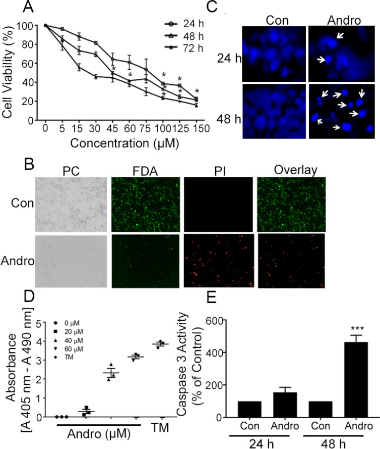
Figure 1. Andrographolide suppresses cell proliferation and cell survival in COLO 205 cells.
(A) Cells were treated with Andro for 24, 48 and 72 h and cell viability was quantified by MTT assay. (B) Fluorescence microscopy images showing the viability of COLO 205 cells cultured in vitro with or without Andro (45 μM) (left to right: phase contrast (PC) image, FDA stain, PI stain, overlay of FDA and PI stain). (C) Cells were treated with Andro IC50 dose (45 μM) for either 24 h (upper panels) or 48 h (lower panels) and stained with DAPI. Apoptotic cells were identified by condensation and fragmentation (arrows) of nuclei using inverted fluorescence microscope. (D) Detection of nucleosomes in cytoplasmic fractions at increasing doses of Andro and TM. 104 cells were treated with or without Andro (20, 40, or 60 μM) or TM (1μg/ml) for 48 h at 37°C. Cell lysates (20 μl) were analyzed in the ELISA. (E) Caspase 3 activity was evaluated by Caspase-3 colorimetric activity assay kit as described. Experiments were performed two times (D and E) or three times (A, B and C). (*P < 0.05, ***P < 0.001).

