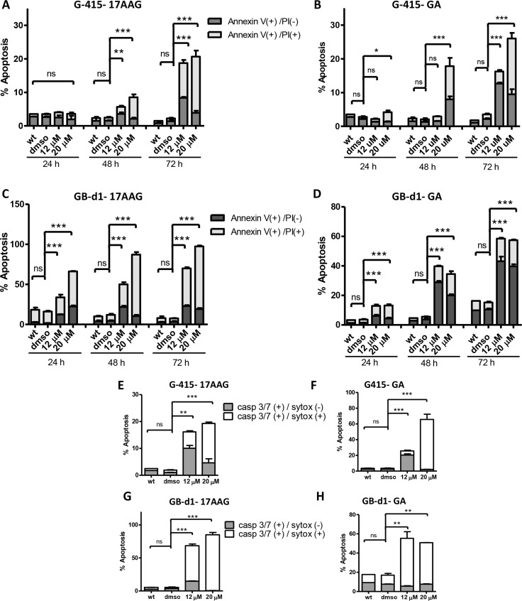
Figure 5. Effect of 17-AAG and GA on apoptosis in human GBC cells.
(A and B) G-415 and (C and D) GB-d1 cells were treated with 17-AAG or GA and harvested after 24, 48, and 72 hours for annexin V /propidium iodide (PI) staining and analized by flow cytometry. Data are shown as mean ± SD (*P < 0.05; **P < 0.01; ***P < 0.001; ns: not significant). Activity of Caspase-3/7 in G-415 cells (E and F) and in GB-d1 cells (G and H) treated with 17-AAG or GA for 72 h was assessed by Flow Cytometry using a fluorogenic substrate for detection of activated caspases 3 and 7 in apoptotic cells. Data are shown as mean ± SD (**P < 0.01; ***P < 0.001; ns: not significant).

