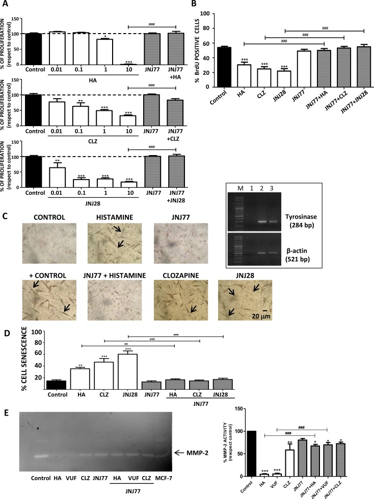
Figure 2. H4R-induced biological responses in 1205Lu cells.
Cells were left untreated (control) or treated with histamine (HA), clozapine (CLZ), JNJ28610244 (JNJ28) or VUF8430 (VUF) and/or 10 μM JNJ7777120 (JNJ77) (A) Clonogenic assay. (B) Incorporation of BrdU-positive cells assessed by immunocytochemistry 48 h after treatment. (C) L-dopa staining with melanin precursor (10 mM L-dopa). evaluated 10 days after treatment. Localization of dopa-oxidase was indicated by the presence of an insoluble brown/black precipitate. Pictures were taken at 200X-fold magnification. Scale bar = 20 μm. Arrows indicate cell prolongations. Positive control: 16 nM TPA + L-dopa. Inset: Tyrosinase expression in 1205Lu was evaluated by RT-PCR (284 bp). Lanes: M, DNA ladder molecular size marker; 1, Negative control (without cDNA); 2: Positive control (M1/15 human melanoma cells), 3: 1205Lu cells. β-actin (521 bp) was used as load control. (D) Senescence-associated to β-galactosidase staining evaluated 48 h after treatment. (E) MMP-2 gelatinololytic activity determined 24 h after treatment, MCF-7 cells were used as positive control. Error bars represent the means ± SEM of three independent experiments (ANOVA and Dunnett's Multiple Comparison Test, *P < 0.05, **P < 0.01, ***P < 0.001 vs. control; ANOVA and Newman–Keuls Multiple Comparison Test, ##P < 0.01, ###P < 0.001 vs. JNJ77+HA or H4R agonist). Results are representative of three independent experiments.

