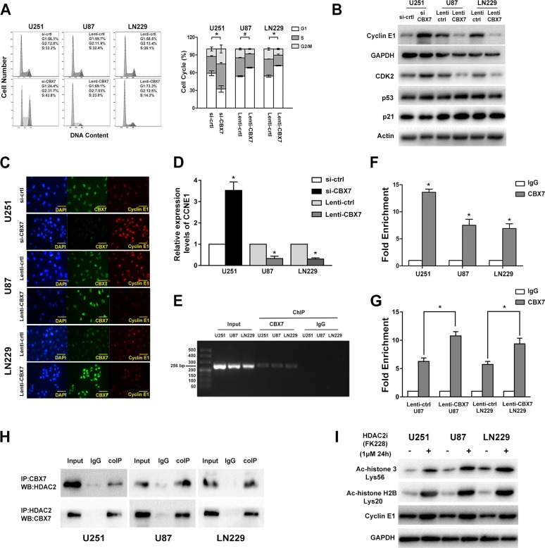
Figure 4. CBX7 induces G1/S arrest through binding and silencing CCNE1 promoter which mediated by the recruit of HDAC2.
(A) Flow cytometic assay was used to assess the cell cycle in GBM cells transfected with siRNAs or lentivirus vectors and the right panel showed the percent of different cell cycle phases. (B) Cyclin E1, CDK2, p53 and p21 levels were tested after the regulation of CBX7 in different GBM cell lines. (C) Immunofluorescence staining of CBX7 (green) and Cyclin E1 (red) in in different GBM cells. Scale bar = 50 mm. (D) Relative expression of CCNE1 in CBX7 up/down regulated cells was studied by Q-PCR. *P < 0.05. (E, F) ChIP experiment was used to examine the interaction between CBX7 and CCNE1 promoter. The precipitate was collected and evaluated by both RT-PCR and q-PCR. *P < 0.05, #P < 0.001. (G) ChIP assay showed that the occupancy of CCNE1 promoter by CBX7 was increased in CBX7 overexpression U87 and LN229 cells. P < 0.05. (H) Co-IP analysis was performed to evaluate the interaction of CBX7 and HDAC2. Total cell lysates from GBM cells were co-immunoprecipitated by anti-CBX7 antibody and anti-HDAC2 antibody. Protein-antibody complexes were analyzed by western blot and the results showed that CBX7 binds to HDAC2 in three cell lines. (I) After treated with 1μM HDCA2 inhibitor FK228 for 24 h, the degree of histone acetylation (acetyl-histone H3 Lys56 and acetyl-histone H2B Lys20) and cyclin E1 levels in three GBM cell lines were tested via western blot.

