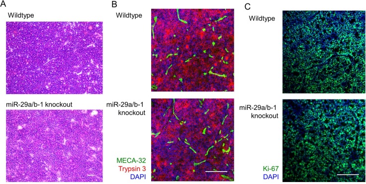
Figure 3. miR-29a-deficient tumours present with normal histopathological growth.
Ela1-TAg+ mice, on the wildtype and miR-29a knockout backgrounds were followed to 21 weeks of age, at which point tumours assessed by fresh frozen histology. (A) H&E histological assessment. Scale = 100 μm. (B) Trypsin 3 (acinar cell carcinoma marker), MECA-32 (vascularisation marker) and DAPI staining by immunofluorescence. Scale = 50 μm. (C) Ki67 (proliferation marker) and DAPI staining by immunofluorescence. Scale = 50 μm. Representative images of n = 3/group displayed.

