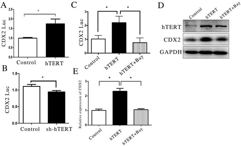
Figure 2. hTERT increase the activity of CDX2 promoter partly through NF-κB signaling pathway.
(A and B) The luciferase activity of CDX2 in MKN45 cells and AGS cells, after hTERT was over-expressed or suppressed, respectively (unpaired t-test was used to analyze data, *p < 0.05). (C) The luciferase activity of CDX2 in MKN45 cells, after the NF-κB signaling pathway was blocked (unpaired t-test was used to analyze data, *p < 0.05). (D and E) The mRNA and protein levels of CDX2 were measured using qRT-PCR and WB, after the NF-KB signaling pathway was blocked (unpaired t-test was used to analyze data, *p < 0.05).

