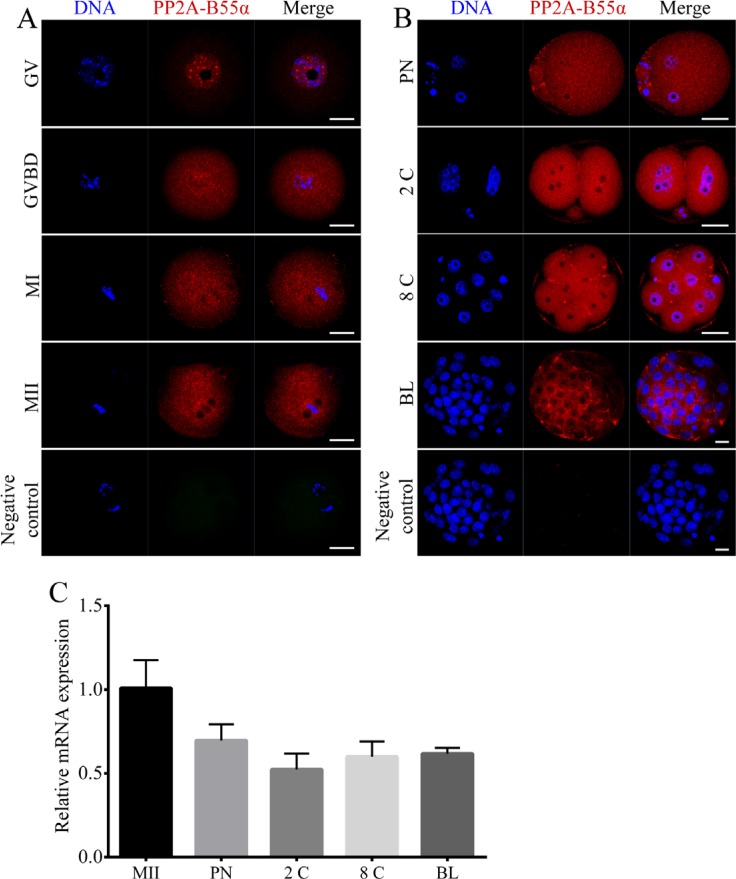
Figure 1. Localization and expression patterns of PP2A-B55α in mouse oocytes and preimplantation embryos.
(A) Subcellular localization of PP2A-B55α from the GV stage to the MII stage of mouse oocyte meiotic maturation. PP2A-B55α mainly localized in the nucleus at the GV stage. After the GVBD stage, PP2A-B55α was distributed throughout the oocyte. (B) Subcellular localization of PP2A-B55α during mouse embryonic development. Blue, DNA; red, PP2A-B55α. Bar = 20 μm. (C) PP2A-B55α transcript levels determined by real-time RT-PCR at different stages of mouse oocyte meiotic maturation and embryonic development. 2C: 2-cell; 8C: 8-cell; BL: blastocyst.

