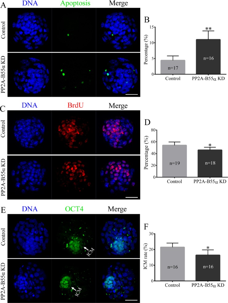
Figure 7. Knock down of PP2A-B55α affects the levels of apoptosis and cell proliferation in mouse embryos.
(A) Representative images of embryos at the blastocyst stage in the TUNEL assay. Bar = 50 μm. (B) The percentage of apoptotic cells in blastocysts that developed in vitro. (C) Immunofluorescence staining of BrdU in mouse embryos at the blastocyst stage. Bar = 50 μm. (D) Percentages of BrdU-positive cells in blastocysts. (E) Immunofluorescence staining of OCT4 in mouse embryos at the blastocyst stage. Bar = 50 μm. (F) ICM rate of blastocysts. The numbers of blastocysts examined in each experimental group are shown in the bars. The data are the mean ± SD of three independent experiments. Statistically significant differences are indicated by asterisks (*p < 0.05, **p < 0.01).

