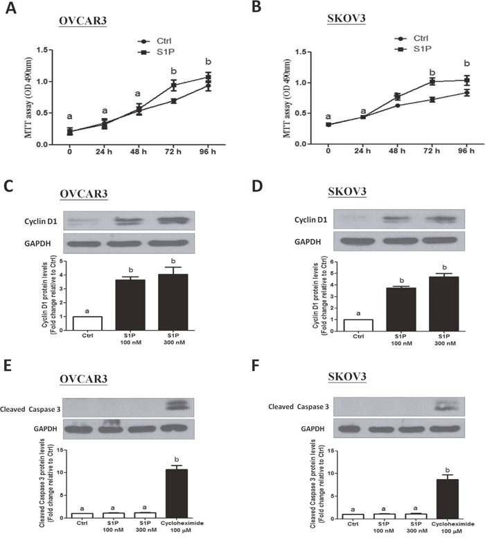
Figure 4. S1P stimulates cell proliferation in OVCAR3 and SKOV3 cells.

(A-B) Cells were seeded in a 96-well plate. After incubation for 24 h, S1P or PET solution (vehicle control) were added for additional 72 h. The cell viability was evaluated using an MTT assay. (C-D) Cells were treated with 100 or 300 nM of S1P for 24 h, and cyclin D1 protein levels were examined using Western blot analysis. (E and F) Cells were treated with S1P (100 or 300 nM) or cycloheximide (100 μM) for 24 h, and the cleaved caspase 3 protein levels were examined using Western blot analysis. The results are expressed as the mean ± SEM from at least three independent experiments. All samples were compared using one-way ANOVA followed by Tukey's multiple comparison tests, and values without a common letter (a, b, c and d) are significantly different (P<0.05).
