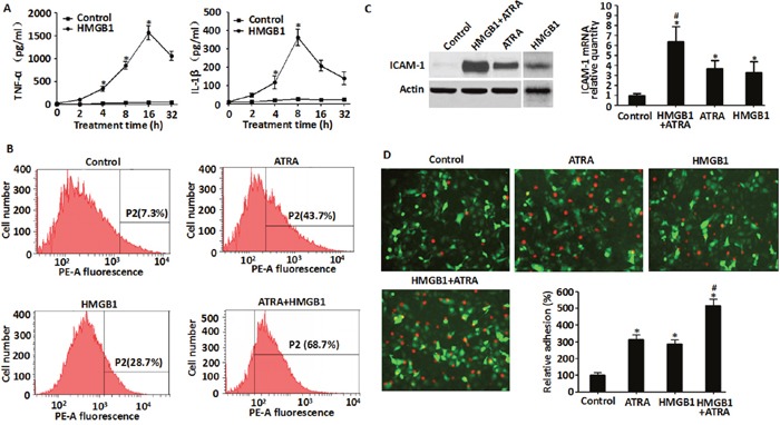
Figure 4. Exogenous HMGB1 induced cytokine secretion, up-regulated expression of ICAM-1 and enhanced endothelial adhesion in NB4 cells.

A. Levels of TNF-α and IL-1β secreted by NB4 cells that were treated with HMGB1 (10 μg/ml) for 2-32 h were detected by ELISA. (n=3, *P<0.05 versus control group). B. and C. Levels of ICAM-1 expressed in NB4 cells that were treated with ATRA (1 μM) for 48 h, HMGB1 (10 μg/ml) for 8 h or ATRA (1 μM) for 48 h followed by HMGB1 (10 μg/ml) for 8 h were determined by flow cytometry, western blot and qRT-PCR. ICAM-1 levels were expressed as a percentage of control (DMSO) (n=3, *P<0.05 versus control group; #P<0.05 versus ATRA or HMGB1 group). D. Determination of cell adhesiveness of NB4 cells after treatment with ATRA (1 μM) for 48 h, HMGB1 (10 μg/ml) for 8 h or ATRA (1 μM) for 48 h followed by HMGB1 (10 μg/ml) for 8 h. Towards this, NB4 cells were fluorescently tagged by CM-Dil. Then, the fluorescent NB4 cells were co-incubated with EA.hy926 endothelial cells (transfected with pEGFP-N1 vector) and observed microscopically (100X magnification) and quantified fluorometrically. The data were expressed as a percentage of the DMSO control (n=3, *P<0.05 versus control group; #P<0.05 versus ATRA or HMGB1 group).
