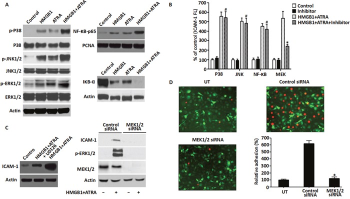
Figure 6. Role of MEK/ERK pathway in HMGB1-mediated cytokine secretion and ICAM-1 elevation.

A. The MAPK signaling pathway was analyzed in NB4 cells that were treated with ATRA (1 μM) for 48 h or/and HMGB1 (10 μg/ml) for 8 h by western blot analysis of phosphorylated and non-phosphorylated p38, Jnk and Erk kinases. Also, NF-kB pathway was analyzed by detecting the levels of p65 and IkB-α proteins for the same treatments described above along with PCNA. Actin was used as control. B. ICAM-1 levels were determined by FACS analysis of NB4 cells that were treated with ATRA (1 μM) for 48 h followed by HMGB1 (10 μg/ml) for 8 h with or without pre-treatment of the p38, Jnk, NF-kB or MEK inhibitors for 30 min (n=3, *P<0.05 versus MEK ATRA+HMGB1 group; #P>0.05 versus P38, JNK or NF-κB ATRA+HMGB1 group). C. The requirement for MEK/ERK signaling in DS was analyzed by detecting the levels of ICAM-1, MEK1/2 and p-ERK by western blot in NB4 cells that after pre-treatment with U0126 (20 μM) for 30 min or transfection with MEK1/2 siRNA for 48 h were induced by ATRA (1μM) for 48h followed by HMGB1 (10 μg/ml) for 8 h. D. Adhesion property of NB4 cells that were transfected with MEK1/2 or control siRNA for 48 h and treated with ATRA (1 μM) for 48 h followed by HMGB1 (10 μg/ml) for 8 h was analyzed as described in the methods. Briefly, the treated cells were fluorescence-labeled with CM-Dil and then co-incubated with EA.hy926 endothelial cells (transfected with pEGFP-N1 vector) to monitor cell-cell adhesion. Fluorescent NB4 cells adherent on endothelial cells were observed microscopically and quantified fluorometrically (100X magnification). The data were expressed as percentages relative to the control group (n=3, *P<0.05 versus control siRNA group).
