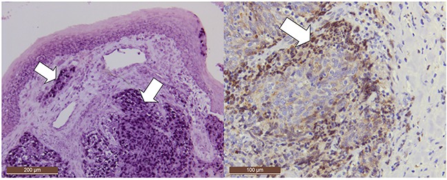
Figure 1. Positive controls: EBER positivity in a nasopharyngeal carcinoma (NPC) with EBER-ISH (deeper purple, mostly nuclear staining, arrows).

EBER is intensively expressed in all NPC cells. Note that EBER is also expressed in localized area of normal surface epithelium. Additionally, LMP-1 was detected by immunohistochemistry in the cancer cells of invasion front in addition to occasional lymphocytes in that region (dark brown, 50x and 100x magnifications, respectively).
