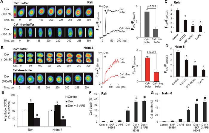
Figure 3. Dexamethasone stimulates intracellular Ca2+ release and SOCE and co-treatment with dexamethasone and SOC inhibitors markedly enhances ALL cells death.

Cells were loaded with Fura-2/AM, and the changes in intracellular Ca2+, [Ca2+]i, (F340/F380) were monitored. (A, B) The colored time-lapse images and the graphical representation show the changes in [Ca2+]i evoked by dexamethasone (Dex), in Reh and Nalm-6 cell lines, respectively, in Ca2+-containing and in Ca2+-free buffer. (C, D) ALL cells before exposure to 100 nM dexamethasone (Dex) in Ca2+ buffer were preincubated (20 minutes) with U73122 (10 μM), SKF96365 (10 μM) or 2-APB (10 μM). Data are mean ± SEM (n = 5). (E) Effect of dexamethasone (Dex) on SOCE activation in ALL cells. ALL cells ER calcium stores were depleted with thapsigargin (TG, 1 μM) in calcium-free suspension medium in the presence or absence of 2-APB (10 μM), cells were then treated without or with 100 nM dexamethasone, followed by addition of 1.8 mM CaCl2. Data are mean ± SEM (n = 3). Reh (F) and Nalm-6 (G) cells were treated with SOC inhibitors (SKF 96365, 5 μM and 2-APB, 5 μM) and dexamethasone (100 nM) alone or in combination for 48 h. Cell death was detected by MTT metabolic colorimetric assay. Data are representative of triplicate experiments. *p <0.001 vs. control; #p <0.001 vs. Dex.
