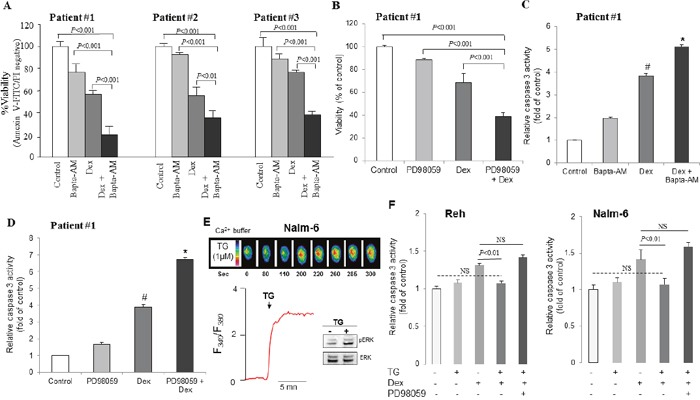
Figure 9. Dexamethasone-induced apoptosis is enhanced by chelating Ca2+ signaling and inhibition of ERK1/2 pathway in primary blasts from ALL patients.

(A) Apoptotic cells were measured by annexin V/FITC-PI staining followed by FACS analysis after 48 hours of treatment in ALL blasts. The percentage of cell viability was calculated by annexin V-FITC negative and PI-negative population and the values of control cells were considered as 100%. (B) Cell viability was detected by MTT metabolic colorimetric assay. The values of control cells were considered as 100%. Data represent the means ±SEM of triplicates. (C, D) Caspase 3 activity was measured with SAFAS Xenius XC Spectrofluorometer. *P<0.01 vs Dex alone treatment; #P<0.05 vs control. Data are mean ± SEM (n = 8). (E) Effect of thapsigargin (TG, 1 μM) on the changes in intracellular Ca2+ and ERK1/2 phosphorylation. (F) Effect of TG (100 nM) on Dex-induced caspase 3 activity. Data are mean ± SEM (n = 3).
