
Figure 2. Bubbles filling the RA and RVOT were detected by TEE in one patient in the S group during hepatic parenchymal resection.
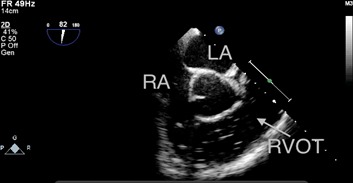
The patient exhibited notable hypotension and a decrease in end-tidal carbon dioxide (from 37 mmHg to 12 mmHg). RA: right atrium, LA: left atrium, RVOT: right ventricular outflow tract.