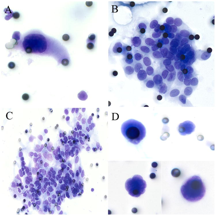
Figure 1.
Cytologic characteristics of circulating CECs. 1A: A paucicellular (<10 cells) specimen consisting of a single markedly enlarged cell with nuclear enlargement (>3× pore size), nuclear hyperchromasia, and nuclear membrane irregularity. 1B: A moderately cellular specimen (10-100 cells) consisting of clusters of epithelioid cells with oval nuclei and occasional nuclear grooves. 1C: A markedly cellular specimen (>100 cells) with clusters of epithelioid cells. 1D: suspicious specimens consisting of markedly enlarged, irregular nuclei but no visible cytoplasm.

