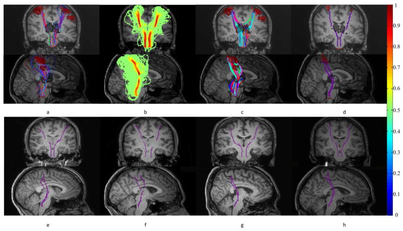
Figure 1.
Tracking and skeletonization of the projection pathways. (a) Probabilistic tracking of fibers from thalamus to S1 (and pons). (b) Probability density of fibers connecting thalamus and S1 (and pons). (c) Streamlines generated by backtracking from S1 (and pons) to thalamus. (d) Skeletons of fiber bundles connecting thalamus and S1 (and pons). First and second rows are coronal and sagittal views respectively. (e–h) Representative skeletons from four other subjects shown in coronal and sagittal views. Note that the colorbar on the far right is for panel (b).

