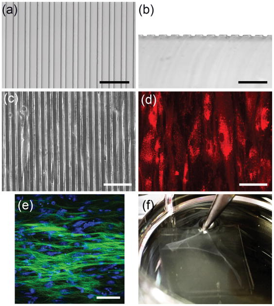
Figure 1. Formation of scaffold-free tissue sheet with parallel cell and matrix organization.
Light micrographs of (a) top view and (b) cross sectional view of the PDMS substrate show grooves approximately 10 μm wide, 10 μm apart, and 5 μm deep. (c) Phase contrast image shows CSSC cultured on the grooved substrate. (e) For better visualization, CSSC were labeled with DiI (red) and cultured on grooved substrate. (e) Two-photon micrograph of 10-day cultures of CSSC on grooved substrates in keratocyte differentiation medium (KDM) shows deposition of parallel organized collagenous matrix (green). Nuclei (blue) were stained by SYTOX-green (blue). (f) After 10 days of culture a robust tissue sheet is formed that can be separated from the substrate using forceps. Scale bars: (a) and (b) = 50 μm, (c)–(e) = 100 μm

